Function of the visual sense
The visual sense system builds a representation of the surrounding environment. It provides humans with the ability to process visual detail, as well as the formation of perception of several non-image related photo-response functions, such as:
- Informing the sleep mechanism (when is it day or night)
- Contributing to the vestibular sense (which way up and round am I)
- Contributing to the proprioceptive sense to coordinate balance, and effectively use motor skills.
The system carries out a number of complex tasks, including:
- Receiving light and forming monocular representations
- Neural mechanisms underlying stereo vision
- Identifying and categorising visual objects
- Assessing distances to and between objects
- Perceiving motion
- Guiding body movements in relation to the objects seen
- Colour vision.
Visual regulation
Much like with auditory regulation, visual regulation means detecting and perceiving the right balance of information from stimulus related to this system. This will be common across the senses. The individuality of this balance is the same as well.
Processing differences
Processing differences in vision can also be present in various submodalities, usually in areas such as to motion, depth perception, colour, brightness and focus. People with differences in their visual perception may:
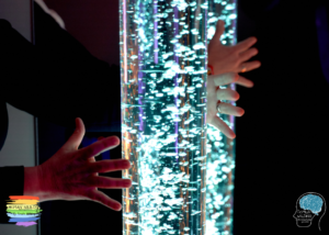
- Stare at spinning objects for long periods
- Spin in circles to gain increased visual feedback
- Wiggle their fingers in front of their face
- Turn away from a subject of their attention (someone talking to them) in order to remove visual distraction and signal detect audition
- Wear sunglasses all year round, and even potentially indoors
- Keep their blinds or curtains closed
- Have highly organised spaces in which they would immediately know if an item has moved
- Complain about double, blurry, or distorted vision in certain circumstances
- Frequently lose their place when reading or copying information
- Not give eye contact, looking beyond someones face, and not being able to focus on anything else if maintaining eye contact.
- Miss visual cues and relies heavily on auditory signals
- Have difficulty identifying specific objects in cluttered spaces, even when focused searching is taking place.
- Frequently misjudge spatial relationships and so may bump into things.
Neurobiology of the visual sense
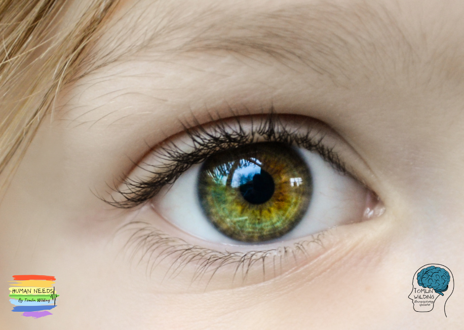
The eye
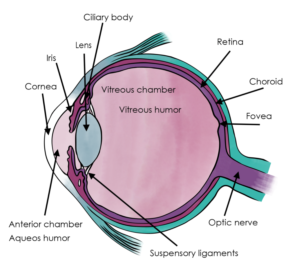
Light is refracted as it enters the eye through the cornea, before passing through the pupil, controlled by the iris, and is then further refracted by the lens. The cornea and lens act together as a compound lens to project an inverted image onto the retina.
The retina consists of a large number of photoreceptor cells, which contain particular protein molecules called opsins. In humans, two types of opsin are involved in conscious vision: rods and cones. There is actually a third type, called melanopsin, though this is not strictly part of the visual system as such, as it serves the body clock mechanism, producing responses in melatonin production.
Rods and cones have different functions. Rods are found primarily around the edges of the retina and are used to facilitate sight at low levels of light, whereas cones are found primarily in the middle of the retina, and are used primarily to distinguish colour, and other features of the visual field at normal levels of light. There are three types of cones that differ in the wavelengths of light they absorb; they are usually called short or blue, middle or green, and long or red.
The optic nerve and chasm
The optic nerves from both eyes meet and cross at the optic chiasm, at the base of the hypothalamus. At this point the data from both eyes in combined and then splits according to the visual field. The corresponding halves of the field of view (right and left) are sent to the left and right halves of the brain respectively, to be processed.
Neurology of vision
The visual sense is processed in the visual cortex, which is the largest system in the human brain and is responsible for processing the visual image. It lies at the rear of the brain above the cerebellum. As visual information passes forward through the visual hierarchy, the complexity of the neural representations increases.
Along with this increasing complexity of neural representation may come a level of specialisation of processing into two distinct pathways: the dorsal stream and the ventral stream, just like in the auditory system. The dorsal stream, commonly referred to as the “where” stream, is involved in spatial attention, and communicates with regions that control eye movements and hand movements. The ventral stream, commonly referred as the “what” stream, is involved in the recognition, identification and categorisation of visual stimuli.